Key Features & Benefits
Clinically relevant – Recapitulates human type I hypersensitivity with IgE-mediated mast cell activation, vasodilation, and pruritus.
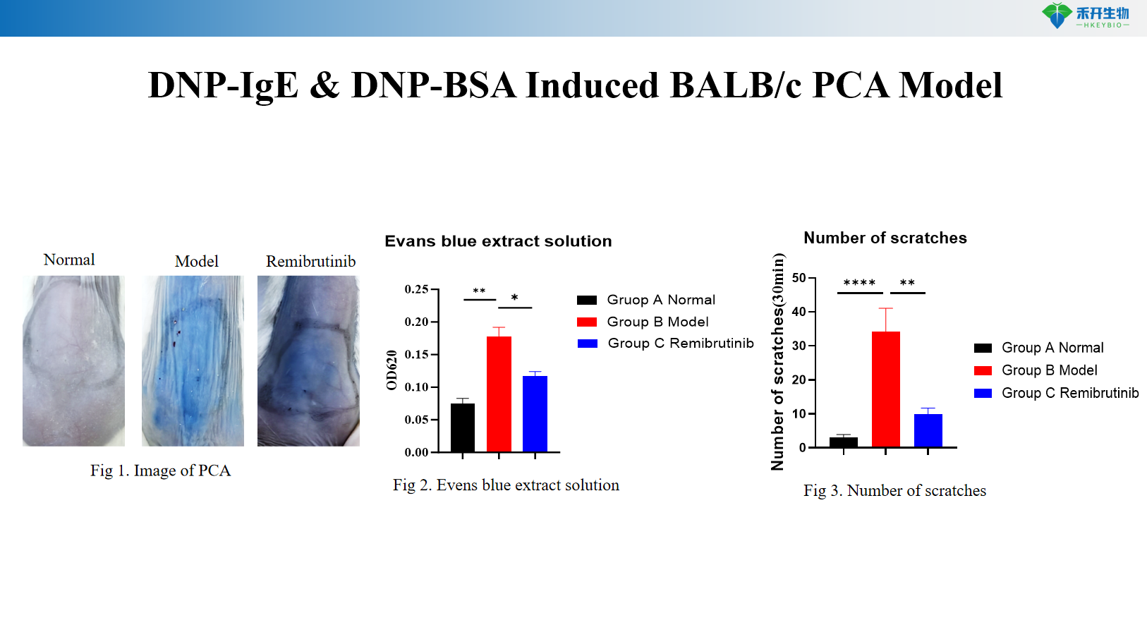
Quantifiable endpoints – Evans blue extravasation (blue spot diameter or OD measurement), scratching behavior count, ear thickness, serum IgE levels.
Two species options – Mouse (BALB/c) and rat (Wistar) models available to suit different experimental needs.
Translational value – Ideal for testing anti-IgE biologics (omalizumab), mast cell stabilizers (cromolyn), H1-antihistamines, and other anti-allergic agents.
IND-ready data packages – Studies can be conducted in accordance with GLP principles.
Technical Data & Validation
DNP-IgE & DNP-BSA Induced BALB/c PCA Model
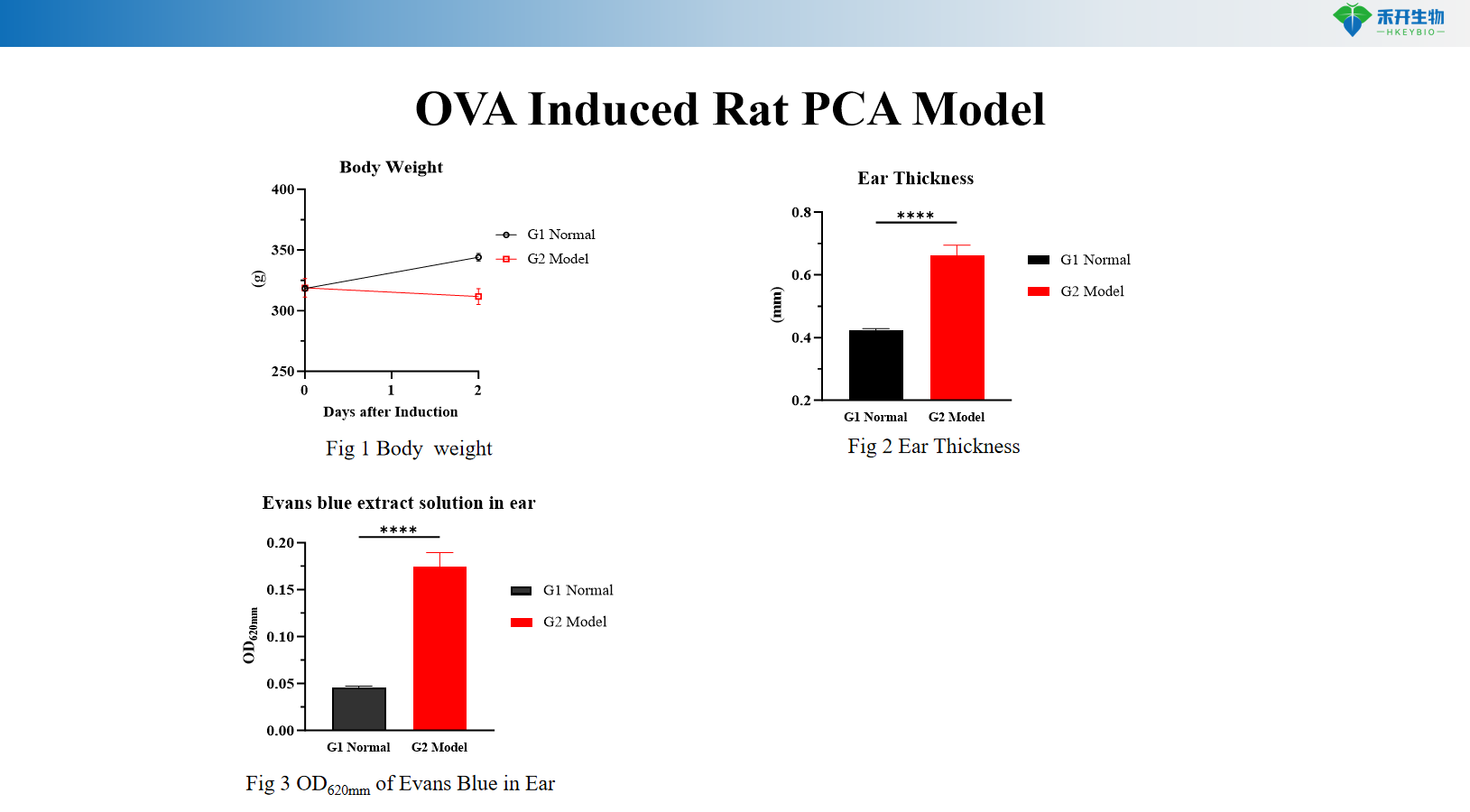
OVA Induced Rat PCA Model
Applications
• Efficacy testing of anti-IgE biologics (omalizumab, ligelizumab) and mast cell stabilizers (cromolyn, ketotifen)
• Evaluation of H1-antihistamines (cetirizine, fexofenadine) and other anti-allergic agents
• Target validation for IgE/FcεRI pathway and mast cell biology
• Biomarker discovery (IgE, histamine, mast cell mediators)
• IND-enabling pharmacology and toxicology studies
Model Specifications
Parameter | Mouse PCA Model | Rat PCA Model |
Species/Strain | BALB/c mouse | Wistar rat |
Induction method | Intradermal injection of DNP-IgE (passive sensitization) + i.v. DNP-BSA with Evans blue | Intradermal injection of OVA-sensitized serum + i.v. OVA with Evans blue |
Study duration | 24–48 hours (sensitization + challenge) | 24–72 hours |
Key endpoints | Evans blue extravasation (blue spot diameter or OD), scratching behavior count | Body weight, ear thickness, Evans blue extravasation (OD 620 nm), serum OVA-specific IgE, skin histopathology (toluidine blue) |
| Positive control | Anti-IgE antibody or antihistamine (e.g., cetirizine) available as reference compounds |
Data package | Raw data, analysis reports, clinical photographs, ELISA results, histology slides, bioinformatics (optional) |
❓ Frequently Asked Questions
Q1: What PCA models do you provide? A1: We offer two classic PCA models: DNP-IgE & DNP-BSA induced model in BALB/c mice, and OVA-induced PCA model in Wistar rats.
Q2: What is the modeling mechanism? A2: Sensitized IgE binds to skin mast cells. Subsequent antigen stimulation triggers mast cell degranulation and release of allergic mediators, increasing vascular permeability and causing typical cutaneous allergic reactions.
Q3: What evaluation indicators are used? A3: We monitor body weight, observe urticaria, scratching events and ear thickness. We detect IgE levels and conduct toluidine blue staining for pathological analysis. Evans blue leakage is also measured quantitatively.
Q4: What is the experimental timeline? A4: For the DNP-related model: DNP-IgE injection at 0 hour, antigen challenge at 24 hours, and detection finishes in 30 minutes. For the OVA model, the whole experiment lasts 48 hours.
Q5: What are the advantages and applications? A5: These models have stable allergic reactions, clear results and reliable reproducibility. They are widely used for efficacy screening and mechanism research of anti-allergy drugs.


