Key Features & Benefits
Clinically relevant – Recapitulates human radiation dermatitis with acute erythema, desquamation, and chronic fibrosis using standardized RTOG scoring.
Mechanism-driven – X-ray induced DNA damage, ROS production, and TGF-β mediated fibrosis mirror human radiotherapy-induced skin injury.
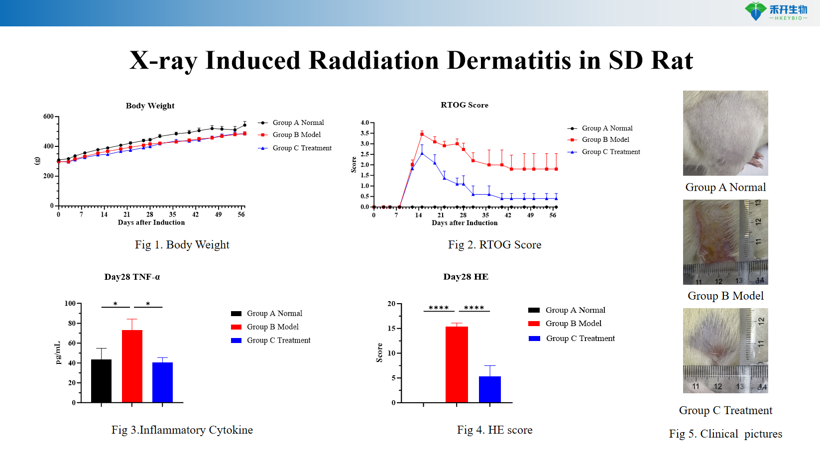
Comprehensive endpoints – Body weight, RTOG clinical score (0–4 scale), serum TNF-α levels, skin histopathology (HE scoring), inflammatory cell infiltration, dermal fibrosis.
Translational value – Ideal for testing radioprotectors, anti-inflammatory agents, antioxidants, and wound healing therapies.
IND-ready data packages – Studies can be conducted in accordance with GLP principles.
Technical Data & Validation
X-ray Induced Raddiation Dermatitis in SD Rat
Applications
• Efficacy testing of radioprotectors (amifostine, superoxide dismutase) and anti-inflammatory agents (corticosteroids, NSAIDs)
• Evaluation of wound healing agents, antioxidants, and TGF-β inhibitors for chronic radiation fibrosis
• Target validation for radiation-induced inflammatory and fibrotic pathways
• Biomarker discovery (TNF-α, TGF-β, oxidative stress markers)
• IND-enabling pharmacology and toxicology studies
Model Specifications
Parameter | Specification |
Species/Strain | Sprague-Dawley rat |
Induction method | Local X-ray irradiation (single dose: 30–50 Gy or fractionated doses) on shaved dorsal skin, typically 2–3 cm² area |
Study duration | Acute: 1–4 weeks; Chronic: 4–12 weeks (for fibrosis studies) |
Key endpoints | Body weight, RTOG clinical score (erythema, dry/moist desquamation, ulceration, fibrosis), serum TNF-α levels (ELISA), skin histopathology (HE staining with scoring for inflammatory infiltration, epidermal thickness, dermal fibrosis), optional: immunohistochemistry (TGF-β, α-SMA, collagen I), oxidative stress markers (MDA, SOD)Body weight, RTOG clinical score (erythema, dry/moist desquamation, ulceration, fibrosis), serum TNF-α levels (ELISA), skin histopathology (HE staining with scoring for inflammatory infiltration, epidermal thickness, dermal fibrosis), optional: immunohistochemistry (TGF-β, α-SMA, collagen I), oxidative stress markers (MDA, SOD) |
| Positive control | Amifostine or corticosteroids available as reference radioprotective/anti-inflammatory compounds |
Data package | Raw data, analysis reports, clinical photographs, RTOG scoring records, ELISA results, histology slides, bioinformatics (optional) |
❓ Frequently Asked Questions
Q1: What radiation dermatitis model do you provide? A1: We offer an X-ray-induced radiation dermatitis model using SD rats for preclinical research on radiation-related skin injury.
Q2: What is the modeling mechanism? A2: X-ray radiation causes DNA damage and reactive oxygen species production in skin cells. It triggers acute inflammation and long-term TGF-β overexpression, leading to skin fibrosis, atrophy and typical radiation dermatitis lesions.
Q3: What evaluation indicators are applied? A3: We monitor body weight and assess skin lesions via RTOG scoring. We also detect TNF-α level and perform H&E staining for pathological analysis.
Q4: What is the experimental cycle? A4: X-ray irradiation is performed on Day 0. The whole experiment lasts 28 days until all observations and tests are completed.
Q5: What are the advantages and applications? A5: This model highly mimics the progression of human acute and chronic radiation dermatitis with stable symptoms. It is widely used for efficacy screening and mechanism research of radioprotective and therapeutic drugs.


