Key Features & Benefits
Clinically relevant – Recapitulates human SSc-ILD with pulmonary fibrosis, skin fibrosis, and TGF-β mediated pathology.
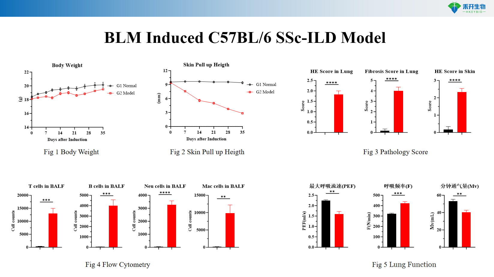
Comprehensive endpoints – Body weight, skin pull-up height (skin fibrosis), lung function, flow cytometry (immune cell infiltration), lung histopathology (HE & Masson staining).
Mechanism-driven – BLM induces DNA damage, oxidative stress, and TGF-β pathway activation, mirroring human SSc-ILD pathogenesis.
Translational value – Ideal for testing anti-fibrotic agents (nintedanib, pirfenidone), TGF-β inhibitors, and immunomodulators.
IND-ready data packages – Studies can be conducted in accordance with GLP principles.
Technical Data & Validation
BLM Induced C57BL/6 SSc-ILD Model
Applications
• Efficacy testing of anti-fibrotic agents (nintedanib, pirfenidone, TGF-β inhibitors, galectin-3 inhibitors)
• Evaluation of immunomodulators targeting inflammation and fibrosis
• Target validation for TGF-β signaling and fibrotic pathways
• Biomarker discovery (collagen markers, inflammatory mediators, immune cell signatures)
• IND-enabling pharmacology and toxicology studies
Model Specifications
Parameter | Specification |
Species/Strain | C57BL/6 mouse |
Induction method | Intratracheal instillation of bleomycin (BLM, 1–2 U/kg) in saline, single dose |
Study duration | 14–28 days (fibrosis development) |
Key endpoints | Body weight, skin pull-up height (skin fibrosis), lung function (compliance, resistance), flow cytometry (immune cell infiltration: macrophages, neutrophils, T cells), lung histopathology (HE and Masson trichrome staining with Ashcroft score), optional: hydroxyproline content, BALF cell counts, cytokine levels |
| Positive control | Nintedanib or pirfenidone available as reference anti-fibrotic compounds |
Data package | Raw data, analysis reports, lung function data, flow cytometry files, histology slides (HE, Masson), bioinformatics (optional) |
❓ Frequently Asked Questions
Q1: What SSc-ILD preclinical model do you offer? A1: We provide a bleomycin (BLM)-induced SSc-ILD model using C57BL/6 mice for related disease research and drug evaluation.
Q2: What is the modeling mechanism? A2: BLM causes DNA damage and oxidative stress in lung tissue. It activates sustained inflammation and abnormal tissue repair, and drives lung fibrosis via the TGF-β pathway, which recapitulates typical pathological features of human SSc-ILD.
Q3: What evaluation indicators are adopted? A3: We monitor body weight and test lung function. We also detect inflammatory cytokines, perform flow cytometry analysis, and conduct HE and Masson staining for lung pathological assessment.
Q4: What is the experimental schedule? A4: BLM is administered on Day 0. Lung function testing is conducted on Day 34, and the whole experiment concludes on Day 35.
Q5: What are the model advantages and applications? A5: This model presents stable inflammation and fibrotic lesions with high similarity to human SSc-ILD. It is widely used for efficacy screening and mechanism study of anti-fibrotic and anti-inflammatory drugs for SSc-ILD treatment.


