Key Features & Benefits
Clinically relevant – Mimics human SSc: skin fibrosis, collagen accumulation, and autoimmune features.
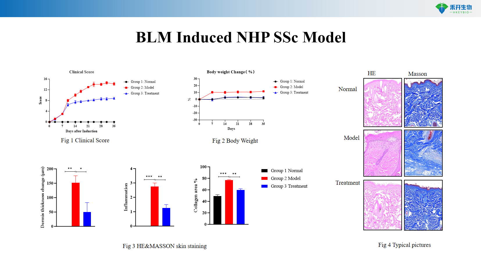
Well-characterized endpoints – Clinical score, body weight, histopathology (H&E, Masson's trichrome), collagen quantification.
Mechanism-driven – BLM induces fibrosis through DNA damage and inflammatory pathways, closely mimicking human disease pathogenesis.
Translational value – Ideal for testing anti-fibrotic agents (TGF-β inhibitors, tyrosine kinase inhibitors), immunomodulators, and biologics.
IND-ready data packages – Studies can be conducted in accordance with GLP principles.
Technical Data & Validation
Representative data from our BLM Induced NHP SSc model:
BLM Induced NHP SSc Model
Applications
• Efficacy testing of anti-fibrotic agents (TGF-β inhibitors, tyrosine kinase inhibitors, pirfenidone, nintedanib)
• Target validation for fibrosis pathways (collagen synthesis, TGF-β signaling)
• Biomarker discovery (collagen metabolites, autoantibodies)
• Mechanism of action (MOA) studies
• IND-enabling toxicology and safety pharmacology studies
Model Specifications
Parameter | Specification |
Species | Cynomolgus macaque (Macaca fascicularis) |
Induction method | Intradermal injections of bleomycin (BLM), multiple sites, repeated dosing for 4–8 weeks |
Study duration | 6–10 weeks (induction + treatment phase) |
Key endpoints | Clinical score (skin thickness, hardness); body weight; histopathology (H&E, Masson's trichrome for collagen deposition); dermal thickness measurement; optional: autoantibody titers, hydroxyproline assay |
Data package | Raw data, analysis reports, histology slides (H&E, Masson), clinical photographs, bioinformatics (optional) |
❓ Frequently Asked Questions
Q1: What NHP systemic sclerosis (SSc) model do you provide, and what induction method is adopted?
A1: We provide a well-validated bleomycin (BLM)-induced SSc model using cynomolgus macaques, which recapitulates core pathological features of human scleroderma.BLM is a copper-chelating peptide that triggers DNA damage and drives progressive dermal fibrosis. Via daily intradermal administration of BLM on dorsal skin, the model develops both robust skin collagen accumulation and autoimmune manifestations including detectable autoantibody production, highly consistent with the pathogenesis of human systemic sclerosis.
Q2: What are the key translational advantages of your NHP SSc model over conventional rodent scleroderma models?
A2: This NHP model delivers significantly higher clinical predictive value for late-stage preclinical development, with core advantages including:
High biological homology: The skin architecture, immune system composition, and fibrotic response pathways of non-human primates are far more similar to humans than rodents, ensuring more reliable efficacy prediction for anti-fibrotic and immunomodulatory drug candidates.
Clinically relevant phenotypes: The model develops progressive skin hardening and thickening that closely mimics cutaneous manifestations in SSc patients, supporting more intuitive and quantitative clinical efficacy assessment.
It serves as a critical translational bridge between rodent screening and clinical trials, effectively reducing the risk of attrition in clinical development.
Q3: What key endpoints are evaluated in your BLM-induced NHP SSc studies?
A3: We provide multi-dimensional evaluation covering clinical phenotype, histopathology and molecular levels to support comprehensive efficacy assessment:
Clinical endpoints: Standardized clinical scoring for skin lesions (erythema, induration, thickening) and longitudinal body weight monitoring to track disease progression and drug safety.
Pathological endpoints: H&E staining for dermal structure and inflammatory infiltration assessment, and Masson’s trichrome staining for quantitative analysis of collagen deposition and fibrosis severity.
Customized assays including autoantibody quantification, cytokine profiling and target biomarker detection are also available upon request.
Q4: What types of therapeutic candidates is this NHP SSc model suitable for evaluating?
A4: The model supports efficacy evaluation across a broad range of SSc drug development pipelines, applicable to both early target validation and late-stage translational research:
Anti-fibrotic agents targeting collagen synthesis and extracellular matrix remodeling;
Immunomodulators and biologics targeting core pathogenic pathways such as TGF-β, PDGF and pro-inflammatory cytokines;
Novel topical or systemic therapies for cutaneous sclerosis and systemic fibrotic manifestations.
Q5: What is the typical study timeline, and do you support customized protocols and IND-enabling studies?
A5: A standard BLM-induced NHP SSc efficacy study typically runs 35 days from induction initiation to study endpoint, covering the full disease induction and treatment observation phase.We fully support customized study design: our scientific team can tailor BLM dosage, administration regimen, treatment schedule and endpoint assay panels based on your drug modality and research objectives. Studies can also be conducted in accordance with GLP principles to deliver audit-ready data packages that support IND submissions to global regulatory agencies.


